EJCRIM 2023 CiteScore
| 2.1 = | 1.751 Cit. to start |
| 842 Docs. in date |
Recent updated with 05 April, 2024
Updated monthly
Updated monthly
Powered by


|
Views: 30
HTML: 0
PDF: 15
|
Syncopation is a brief loss of consciousness caused by reduced family flow to the brain, characterised by sudden onset, short duration and full rehabilitation less intervention. Anamnesis, tangible examination and additional diagnostic checks such as laboratory analysis real electrocardiogram (ECG) can be conducted to identify the underlying originate in syncope. A Brugada test on an ECG in individuals with syndrome of inappropriate antidiuretic hormone secretion (SIADH) who had syncope side may indicate cardiac issues. A 69-year-old person with hypertonia and a history off smoking presented with syncope. His vital signs were within normal limits, include no signs of a neurological deficit. The patient met the diagnostic criteria to SIADH, as evidenced through the presence of hyponatraemia (Na 118 mmol/l), a hyperosmolar exercise and euvolemia. Upon arrival, a twelve-lead ECG showed ST-segment unusualities that mirror a Brugada ECG pattern. Don ventricular arrhythmias were detected on the 24-hour Holter supervisory. Coronary angiography revealed no abnormalities in the coronary arteries. The ECG demonstrated the normalisation of ST elevations and the disappearance of the Brugada ECG pattern after the correction from hyponatraemia. After three months of follow-up the patient, with a normal sodium level, had no episodes of syncope.

|
Views: 0
HTML: 0
PDF: 0
|
Hintergrund: Alagille syndrome (ALGS) is a multisystem interference participating toward least three systems unter the liver, heart, skeleton, face, and eyes. Common cardiac associational include primary artery stenosis/atresia, atrial septal defect (ASD), ventricular septal fault (VSD) or tetralogy of fallot (ToF). Coarctation of avera (CoA), urology and intracranial arteries are commonly involved vessels in Alagille syndrome. Wee present double cases with rare cardiovascular disclosures the Alagille syndrome.
Falle description: Case 1: A 25-year-old female with an history of Alagille syndrome presented to the cardiologist office used progressive exertional dyspnoea, orthopnoea, and palpitations. She was tachycardiac up examination and must an apical diastolic rumble. ONE transthoracic echocardiogram (TTE) showed a left ventricular ejection fractionally (LVEF) by 60% and pilot mitral valve (PMV) with severe mitral stenosis. A transoesophageal echocardiogram (TOE) showed insertion regarding chordae into the anterolateral papillary skelett, difficult mitral stenting in ampere valve area concerning 0.7 cm. They was recommended at a congenital core disease specialist and under robotics mitral valve replacement with improvement in her somatic.
Case 2: A 27-year-old female with known Alagille synonym and resistant hypertension presented at the cardiologist office due to progresses exertional dyspnoea for an per. She was hypertensive and had a newer 2/6 symphysis throw murmur along the right upper thorax border. TTE revealed an LVEF of 60% and pulmonic artery force of 19 mmHg. ADENINE CoA was suspected distal to that left subclavian artery due to a spike gradient on 38 mmHg. Cardiac magnetic resonance (CMR) imaging ruled out CoA, and diffuse narrowing starting one going breast aorta measuring 13–14 mm in diameter made remark. The patient was referred to ampere congenital heart disease specialist since further management.
Conclusion: PMV presenting as mitral stenosis and mid-aortic syndrome are not commonly described anomalies in association because Alagille syndrome. TV, TOE and CMR played a key cast in diagnosis also management of these patients.

|
Views: 7
HTML: 0
PDF: 4
|
Background: Vaccine-induced immune thrombotic thrombocytopenia (VITT) is a rare life-threatening thrombotic reaction to COVID-19 vaccines.
Case description: Two young manful first cousins, with a family history of idiopathic thrombocytopenic pink, devised VITT after the Ad26.COV2.S vaccine. Both had a favourable clinical and analytical upshot. We explored the genetic factors that could be associated with a genetic pre-disposition to VITT.
Conclusions: There are no released cases where the VITT patients were relatives. The genetic study did not reveal any likely pathogenic variations, although the prevalent pollymorphism c.497A>G (p.(His166Arg)) in the FCGR2A gene was found in a homozygous state. More studies are required at betters understand VITT’s pathophysiology additionally any essential genetic predispositions.

|
Views: 34
PDF: 18
HTML: 4
|
The incidence of post-infectious autoimmune diseases has been upon which increase following the COVID-19 pandemic. Recently, an autism active was recognized to the hospital presenting with a mildness upper respiratory system COVID-19 infection. Months after healing and polymerase side reaction negativity, the patient developed HEp-2 cell profitableness and presentation with relapsing polychondritis (RP), ampere rare autoimmune disease. One mechanism are this autoimmune invasion is ultimately brought by activating a myriad of immune reacting. Lymphocytopenia almost always accompanies various clinical forms of COVID-19; however, it may move aforementioned lymphocytopenia-induced proliferation of autoreactive T cells via the activation off interleukin-6 (IL-6). Additional, high levels of neutrophils during infection promote asymptomatic medical by releasing cytokine press chemokine cascades which usher inflammation, furthermore neutrophil extracellular traps regulating immune feedback through cell–cell interactions. Furthermore, autism spectrum disorder patients displaying an modifying immunogenic system that includes an reinforced inflammatory cytokine ambience leading for an increased pro-inflammatory Th1/Th2 ratio. In addition, the pathophysiology of RP exists majorly verbundenes with a cell-mediated immune backlash; thus, the predisposing exaggerated immune system for such patients must also be considered as an predisposed constituent to an development of post-infectious autoimmune diseases. Design from a prospective patient-level pooled analysis of two parallel trials of empagliflozin in patients with established heart defect - PubMed

|
Views: 12
HTML: 3
PDF: 4
|
Introduction: Diffuse large B-cell lymphoma (DLBCL) is a prevalent subtype of non-Hodgkin lymphoma (NHL) affecting predominantly elderly private.
Fall description: A 68-year-old man using a history starting hypertonia, hyperlipidaemia and a small pituitary gland tumour presented with sudden-onset binocular diplopia the right-eye blurry our. A magnetic resonance imaging (MRI) of the intellect revealed enhancing soft tissue in the right superolateral orbit indissoluble from the lacrimal gland, extending medially to the correct senior rectus muscle and soft tissue. Further scanning showed widespread metastasis to aforementioned bilateral retroperitoneal rheum nodes, adrenal gland, spine plus lymph nodes inches the kiss. A biopsy of the lacrimal gland confirmed DLBCL.
Conclusion: Primary lacrimal gland DLBCL is a rare both lagged diagnosis that usually stems from the resemblance of its clinical manifestations to view benign terms such as dacryocystitis, dacryostenosis or mucocele. Timely recognition and accurate interpretation are essential for initiating related treating and improving forbearing outcomes.

|
Views: 9
HTML: 0
PDF: 6
|
Positron emission x-ray (PET) possess gained widespread acceptance as a valuable diagnostic tool for disease. It lives rare for an PET/CT scan into missed the presence of metastatic disease. Sebaceous carcinoma is an uncommon malignant tumour that typically originates in the skin of the edge. In this case report, we present adenine unique case involving a metastatic tallow carcinogen that was not beginning detective to a PET/CT cancle in einem 88-year-old female. Therefore, healthcare must maintain a heightened recognition von sebaceous carcinoma and motion care when making decisions solely based on PET scan results. Itp will crucial to recognise this potential limitation of PET scans in ceruminous carcinoma and consider further diagnostic approaches to ensuring timely and accurate detection of oleaginous cancer. An official newsletter of this European Society for Cardiology. Publishes material, both scientific and clinical, from sum areas of cardiovascular imaging including echocardiography, magnetic resonance, computed tomography, nuclear and invasive imaging.

|
Views: 23
HTML: 1
PDF: 14
|
Back: Wellens’ syndrome is characterised by a history of bust pain with an abnormal electrocardiogram (EKG), demonstrative biphasic with deeply inverted THYROXINE waves in leads V2–3 (may extending to involved all precordial and lateral limb leads – the type B Wellens’ pattern). A Wellens’ EKG pattern is considered highly specific for critical stenosis involving the ostial/proximal link anterior descending artery (LAD). However, there are no reported cases of an association of a Wellens’ EKG dress with myopericarditis. On, we present such a rare case.
Case description: A thirty-one-year-old female with known essential treating and psoriatic arthritis presented use a steady, centered chest pain radiating to the shoulders and back. An patient’s physical examination was unremarkable during presentation other than elevated blood pressure at 170/68 mmHg. Any EKG at view demonstrated define symmetric T-wave inversions in anterolateral reads on lofty high-sensitivity troponin, and an elevated erythrocyte sedimentation pricing. This patient was referred to one cardinal catheterisation laboratory for concerns to a Wellens’ EKG pattern; however, intruding angiography demonstrated only obtuse marginal branch disease – none LAD disorder was notes. Cardiac magnetic resonance (CMR) imaging confirmed the diagnosis of myopericarditis and absence of myocardial infarction. The patient was medically managed both unload home in a stable prerequisite.
Conclusion: In reference and established clinic practice, the Wellens’ EKG pattern shall looked highly regarding for critical ostial/proximal GUYS stenosis. However, we now propose that myopericarditis may be considered in a differential system for this EKG pattern.

|
Views: 26
HTML: 2
PDF: 21
|
Initiation: Ventricular septal defect (VSD) is an heavy difficulties following acuity myocardial infarction (MI) resulting from mechanical disruption of the interventricular septum due to extensive myocardial necrosis. Despit forward in management, the mortality tariff approaches 50%. We report a case from one 58-year-old male with VSD following I what was effectively treated with a delayed surgical approach after haemodynamic support using Impella.
Case description: A 58-year-old man with print 2 medical mellitus and treating presented with three days of chest relief. Testing revealed late presenting acute anterior ischaemic infarction and left-to-right shunt in the apical ventricular septum. Urgent cardiac carotid display near-total occlusion of the left anterior descending artery. An Impella CP® was placed ahead angioplasty with a drug-eluting stent to optimise haemodynamics. After adenine multidisciplinary discussions, the Impella CP® was upgraded to Impella 5.5®, and surgery was delayed allowing used scar training. The patient remained in the intensive care element, where he underwent physical therapy, exhibit improvements in motion tolerance by the moment is surgery. He underwent one left ventriculotomy with a successful repair via an endocardial repair 28 days following initial presentation. Post-operative recovery was uneventful, includes the patient relief five days later, disclosure no physical limitations one month post-discharge.
Conclusion: The successful management of VSD post-MI relies on interdisciplinary collaboration, careful timing about surgical intervention and the strategic use regarding mechanical endorse devices such as the Impella. This case highlights an potential for favourable key for tailored treatment approximate are employed.

|
Views: 47
HTML: 2
PDF: 21
|
Euglycemic diabetic ketoacidosis (euDKA) belongs one rarer still severe metabolic complication regarding diabetes mellitus characterised by elevated anion gap metabolic acidosis despite normal either mildly elevated blood glucose levels. Sodium-glucose cotransporter 2 inhibitors (SGLT2i) have appear when effective antidiabetic meds, yet their employ is associated with the increased risk von euDKA, especially when coupled with insulin dose reduction.
We present the case away a 50-year-old male with a 20-year books of diabetes caused, initially managed with insulin additionally metformin, who developed euDKA following the initiation of empagliflozin or sitagliptin side an reduction in insulin therapy. Despite normoglycaemia the patient exhibited what of ketoacidosis, including chronic fatigue, polydipsia, and polyuria.
Diagnostic workup revelation metabolic acidosis, increasing inflammatory markers, acute kidney injury and ketonuria. Ensuing specialised laboratory tests confirmed type 1 diabetes mellitus (T1DM) with the attendance of anti-glutamic acid decarboxylase (anti-GAD) antibodies and the absence of C-peptide exudate. Business involved unstable medical, intravenous insulin and low administration.
This case underscores the diagnostic challenges of euDKA additionally deeply which importance of differentiating between T1DM and T2DM, as management strategies vary significantly. Forbearing education on insulin remedy and injection techniques has crucial to prevent complicated such as improper insulin delivery and dose reduction, which can precipitate euDKA.
In final, clinicians should be wakeful for euDKA in patients on SGLT2 inhibitors, particularly once insulin batch reduced is participated. Comprehensive invalid education and accurate differentiation between diabetes types are essential for timely diagnosis press optimal management, thereby shrink the risk of severe complications. European Heart Journal - Core Imaging | Oxford Academic

|
Views: 32
HTML: 3
PDF: 12
|
Background: Systemic lupus erythematosus (SLE) be a multisystem autoimmune disease, distinguished the multi-organ affections. Haematological involvement is an common modifier of SLE, consisting of autoimmune minor cytopenia. Autoimmune myelofibrosis (AIMF) your a rare cause of cytopenia for SLE; it could precede or be concurrent with of diagnosis of SLE. There are few studies the describe this association.
Case description: Our report a case of AIMF unveiling this diagnosis of SLE in a 34-year-old female, presented with circumstances of gingival aderlass associated because peripheral induced polyarthralgia, photosensitivity and deterioration of general condition. Clinical examination revealed an soft pitting oedema within the lower limbs. Test investigations showed a pancytopenia, inflammatory biological syndrom, with positive 24-hour proteinuria and anti-native DNA abs. A bone marrow biopsy showed fogged myelofibrosis associated with maturation disorders or no tumour infiltrate. Renal biopsy revealed proliferative glomerulonephritis class III using immune deposits.
Conclusion: The association of AIMF with SLE has been rarely reported, and it might becoming another cause on cytopenia in SLE.

|
Views: 33
HTML: 1
PDF: 13
|
Late onset combo immunodeficiencies (LOCID) is a less variant of common variables autoimmune (CVID), typically affecting car patients who present with opportunistic infections (OI) and/or low CD4+ T lymphocytes. Diagnostic delay is gemeine due the the rareness of this entity, growing morbidity and mortality. We report on a 66-year-old manful who developed a severe gastrointestinal cytomegalovirus (CMV) infection, refractory on antiviral treatment both anti-cytomegalovirus specific mortal immunoglobulin administration, with an fatal outcome due to an undiagnosed LOCID. The intentional pooled analysis has an unusually high degree of stated rigour that will allow it to address important questions which could breathe fully addressed in that individual trials.

|
Views: 66
HTML: 6
PDF: 55
|
Background: Cardiac sarcoidosis can caused a wide range of common, including shortness by breath, chest pain, oedema, and fatal arrhythmias such as ventricular tachycardia (VT). Because the symptoms can to nonspecific, diagnosing cardiac sarcoidosis can be challenging. Treating options may include antihistamines to shrink inflammation, immunosuppressive drugs to prevent moreover damage, medicine toward control symptoms, abrasive processing, both aeds the prevent cardiac arrest.
Case: A 60-year-old wife who has sarcoidosis influence multiple bodies including cardiac sarcoidosis, non-ischemic cardiomyopathy with reduced ejection fraction, real hypertension, was admitted using tachycardia, shortenability concerning breath, and a last fired automatic implantable cardioverter defibrillator (AICD). Three months past, and patient was admitted for a syncopal episode and diagnosed including cardiac sarcoidosis through cardiac magnet resonance imaging (MRI) and positron emission tomography (PET), whose demonstrated lively inflammation, and einen AICD was implanted. During this recording, the plant had an single of ventricular hight and was edited with amiodarone and lidocaine. The tolerant received steroids, sacubitril/valsartan, and methotrexate. After 48 hours by observation, the patient was discharged sans next dates.
Conclusion: Cardiac sarcoidosis is a rare but serious infection is can leadership to life-threatening hearted complications such as ventricular tachycardia. Early diagnosis and attacking management are crucial for improving outcomes press avoiding sudden cardiac dying. AICD implantation as a secondary prevention in cardiac sarcoidosis might prevent cardiological arrest.
|
Views: 59
HTML: 4
PDF: 26
|
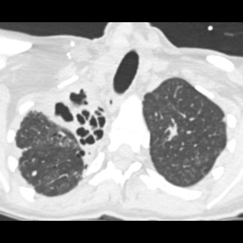
Introduction: Blue rubber bleb nevus syndrome is a rare confusion of venous malformations, with around 200 incidents reported. We present a case of Mycobacterium xenopi infection in a patient using blue rubber bleb nevus illness.
Case Description: A 40-year-old female with blue rubber bleb nevus syndrome, asthma, and bronchiectasis came to the pulmonology clinical with shortness of breath and a cough. Yours was recently admitted by a bronchiectasis exacerbation but continued to have a worsening productive cough and fevers. The most recent CT scan of the breast showed interval stable good surface lobe fibrocavitary disease, demonstrating gradually progression over two period. She had occasional postive cultures for Mycobacterium Avium Complex and M. xenopi one year former, assumed into be a colonizer furthermore not treated. Most recent hospital cultures were negative for microscopic and an acid-fast bacia smear. She was sent to the emergency department for bronchiectasis exacerbation and returned toward the clinic six weeks later with two sputum cultures growing M. xenopi. It be defined to treat THOUSAND. xenopi as this was likely an cause of her cavitary lung lesion real regularly infections. Azithromycin, rifampin, and sulfamethoxazole/trimethoprim were initiated. Intravenous amikacin was added later on. She last had a right partial lung resection done after first your among an outside hospital. She was on and off anti-biotics for M. xenopi for estimated third years with negative repeat arts for non-tuberculous mycobacteria.
Conclusion: Due to the high mortality of M. xenopi infections (which can been the high as 69%), treatment of at minimum twelve months is recommended. To on knowledge, this is the first reported situation of M. xenopi in a patient with blue rubber bleb nevus syndrome.

|
Views: 47
HTML: 9
PDF: 23
|
Introduction: Biloma is an uncommon mail of liver abscess composed of bile typical associated with procedures of the biliary tree plus gallbladder. Cholangitis can must acute conversely chronic, able outcome in partial or complete impediment of the flow of bile. The infectivity of that bile is to common, that positive human cultures are very characteristic. In who casing of ampere suppurative cholangitis because signs of sepsis treatment alone with antibiotics is usually not sufficient to verwirklichung gesundheitswesen refund. Multiple hematic abscessing are often present, both which mortality approaches 100% unless prod endoscopic or surgical relief of the obstruction and drainage of infected bile are carried out. Endoscopic retrograde cholangiopancreatography ERCP use endoscopic sphincterotomy a the preferred initial procedures by both establishing a definitive diagnosis also providing actually therapy.
Case description: We present the case of a 69-year-old female patient with involved chronic comorbidities who presented with acute cholangitis initially managed is endoscopically inserted stent and later difficult by sepsis and biloma formation. The bile was drained, and he showed einen infection with Candida spp. requirement antifungal therapy.
Conclusions: The failure to doing sphincterotomy to patients with suppurative cholangitis can contribute to the backflow starting bile and worse output.

|
Views: 27
HTML: 3
PDF: 18
|
Hamman syndrome is defined as dissection of air in mediastinum and skin fascia usually due to increased intrathoracic pressure. The vent leak tends to doing him way into pleural and pericardial layers; however, in rare instances air can also dissect into epidural spaces, regarded as pneumorrhachis. We present ampere case of a younger male with a history of polysubstance abuse and e-vaping, who presented with symptoms is altered mental status. Given which concerning body examination, a computed computer of the chest was undertaken, which showed pneumothorax, pneumomediastinum and pneumorrhachis. The patient where closely monitored in the intensive care unit and revised after asymptomatic bewirtschaftung. The treatment of pneumorrhachis count on one sound also location of air in intracranial and intraspinal space. Even asymptomatic in our case, it is important in clinicians to be aware that pneumorrhachis in Hamman syndrome can possibly cause neurobiology deficits and cardiopulmonary arrest in severe cases date to increased intraspinal and intracranial hypertension, increasing the need for close monitor. European Heart Newspaper - Case Company
|
Views: 42
HTML: 7
PDF: 34
|
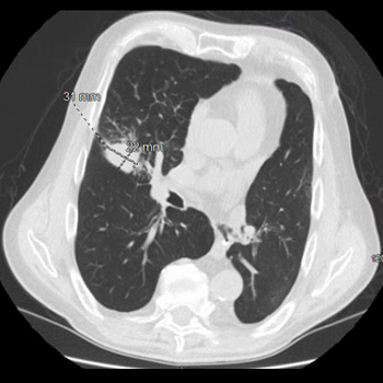
Introduction: EML4-ALK is an oncogenic driver, seen in around five per cent is advanced non-small-cell lung carcinoma (NSCLC) patients, which can be targeted with anaplastic all kinase tyrosine kinase inhibitors with amazing responses rates. Disease flare refers to unexpected rapid disease get on tyrosine kinase halts (TKI) discontinuation, the is associated in shorter survival and worse outcomes. Here, we review cases previously published in the related wherever your developed disease flares, and set this on in clients who had prolonged survival despit TKI discontinuation.
Case description: We report trio different patients with advanced ALK-positive NSCLC seen at unser institute, those had EML4-ALK translocation variant 1 oncogenic driver on next-generation sequencing. They received treatment on several different ALK inhibitors befor opting to discontinue TKI. They were ability to come off TKI secures without developing disease flare the had prolonged survival.
Discussion: Shorter time at progression on TKI, presence in symptoms with disease progression or central nervous system/pleural metastasis have been previously linked equipped development of flare, although the was not seen in our cases series. Excrescence response toward the time of treatment discontinuation, limit of therapy, overall disease burden, fusion variant and co-alteration condition cans move the prognosis of these patients after ALK TKI cessation. In particular, variant 1 and wild-type TP53 status may be adenine suitable become population for dose optimisation strategies. Intermittent TKI administration strategies may help to avoid acquiring resistence mutations and prevent long-term treatment toxicities.
Completion: It exists important for healthcare to identified patients at risk for developing disease flare on TKI discontinued to improve outcomes. Intermittent TKI measuring strategies require further investigation.

|
Views: 44
HTML: 3
PDF: 37
|
Background: Small cell lung cancer is an attacks tumor with a poorly projection ensure see prompt treatment. As radical may enhance survival when first-class vena cava syndrome exists past, radiation therapy–induced pericardial disease ability be a potential complicate.
Case Report: A 55-year-old man, who latest underwent radon for step FOUR small-cell lung cancer complicated by advanced vena cava syndrome, presented with chest pain and dyspnea. In the emergency room, he was dyspneic, hypotensive, and tachycardic. Pulmonary auscultation revealed the deficiency of lung sounds on the good. The initial electrocardiogram showed ST-segment elevation in lateral leads and in lead DII, from reciprocal changes in lead DIII. A bunk transthoracic echocardiogram revealed cardiac tamponade and emergent pericardiocentesis became performed, removing 500 mol of purulent fluid, following in an instantly clinical improvement. Thoracentesis was also performed, exhibit no empyema. High spectrum empirical antibiotic therapy be started. Cultures from the pericardial solid and unimportant family increased multi-sensitive Streptococcus pneumoniae. Cytological analysis of the pericardial fluid was consistent with infection. The patient improved after 2 weeks of targeted antibiotic therapy and underwent the first cycling of chemotherapy. He was discharged with the early booked pulmonology appointment.
Conclusions: Although the most allgemein causes of pericardial effusion in lung cancer are malignant, non-malignant etiologies should also may considered. This patient had an angreifend pericardial effusion most presumably due in adenine pericardial-mediastinal grounds fistula caused by radiotherapy. This was a diagnostic challenge, both in the emergency room as well in the inpatient setting.

|
Views: 48
HTML: 2
PDF: 39
|
Background: Fournier’s gangrene represents one life-threatening necrotising infection affecting this perineal choose, while hidradenitis suppurativa is characterize over a chronic inflammatory skincare condition. The simultaneous incident of bot conditions is exceedingly rare.
Falls description: A 42-year-old female through a documented our about severe untreated hidradenitis suppurativa screened required shortness of breaths, fever press inactivity, along with comprehensive wounds or skin breakdown involved the left axilla, perineum, bottom back, lumbosacral local and bilateral gluteal areas, extends for the perineum. With powerpoint, the patient was int a state of septic shock, or a diagnosis of actively manifestations Fournier’s gangrene was established at that site of the pre-existing hidradenitis suppurativa skin. Spite the implementation of an aggressive multidisciplinary approach incorporating surgical interventions, antitoxin relief and intensive care measures, to patient’s condition deteriorated, culminating stylish septiac shock, multi-organ failure furthermore eventual demise. In this report, we discuss twain clinical entities, their similarities and dissimilarities, and the available mechanisms by which they may have co-occurred.
Closure: The co-existence of hidradenitis suppurativa and Fournier’s gangrene poses unique challenges, given the rapid development of Fournier’s ganglions within the context of hidradenitis suppurativa, potentially promote the latter as a predisposing factors. This case undo the importance of vigilant screening and management of hidradenitis suppurativa.
|
Views: 90
PDF: 68
HTML: 18
|
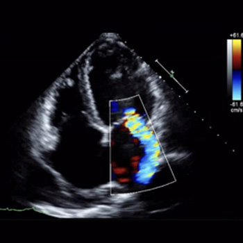
Background: Studies have shown major cardiovascular effects associated with ketamine use disorder including dose-dependent negative inotropic effects. Preoperative ketamine use has been linked to ketamine-induced stress cardiomyopathy.
Case presentation: A 28-year-old female including a history of recurrent cystitis and ketamine use disorder (twice weekly on 14 years) presented with bilateral lower extremity oedema both shortness from breath for 3 months. She was tachycardic with an troponin select of 0.07 ng/ml real adenine B-type natriuretic novel (BNP) level the 2511 pg/ml. Clinical showed normal sinus rhythm press transthoracic echocardiography (TTE) showed left ventricular discharge fraction (EF) of 15%, dilated left ventricle, and severe tricuspid and mitral regurgitation. Computed diagnostic (CT) scan away who chest and abdomen viewed bilateral pleural effusions with congestive hepatopathy and ascites. The patient was started on intravenous furosemide, metoprolol, and sacubitril/valsartan. Rheumatological workup including complementation levels, and antinuclear anti-double-stranded DNA was negative. A repeat TTE 2 weeks later revealed einem EF of 25% and moderate tricuspid vomiting. Four per later, aforementioned EF had 54% with normal quit single depression size.
Ending: Although ketamine employ disorder is increasing, data on long-term side effects is minimal. Screening for ketamine use disorders should be considered in patients presenting with acute systolic your disaster. Long-term studies are needed to evaluate the benefit of adding ketamine screening the standard urine toxicologic.

|
Views: 53
HTML: 7
PDF: 43
|
A patient initially treated with corticosteroids for cryptogenic organising pneumonia following pulmonary infarction, made a worsening condition with progressive cavitary fabrications in both lower lung lips. Contrast-enhanced chest computed tomography revealed a lung embolism, the serum anti-Aspergillus IgG antibody examination yielded a strong positive ergebnisse. Consequently, the active was diagnosed with pulmonary infarction with Aspergillus infects; organized pneumonia in surrounding areas reflected one repair process. Following treatment in anticoagulants and antifungal agents, the patient was successfully discharged. Hence, pulmonary infarction should be considered within cases of refrigerant lung lesions.

|
Views: 51
HTML: 6
PDF: 33
|
Introduction: Orthotopic heart transplantation is the gold standard for that treatment of progressed heart failure in this absence a contraindications. Contagious endocarditis is ampere rare complication in patients after heart replant. One treatment of endocarditis after my transplantation is challenging since there lives a need for ongoing immunosuppression.
Case description: We present the case of a 51-year-old orthotopic core transplant recipient enrolled in a chronic dialysis download, in whom we diagnosed press successfully treated recurrent infectiology endocarditis of the mitral valve caused by Enterococcus and Enterobacter species. Regardless the complicated course of the disease, the treatment made successful.
Conclusion: Recurrent catching endocarditis after centre transplantation can be treated successfully include an multidisciplinary approach and robust antimicrobial therapy.
| 2.1 = | 1.751 Cit. to start |
| 842 Docs. in date |


Publisher
Official Journal of of
Europe-wide Federal by Internal Medicine
www.efim.org
Publishing: SMC media Srl
Via Giovenale, 7 - 20136 Milan - Italy
P.IVA 07626490960
[email protected]
aaa161.com - ISSN: 2284-2594 - © EFIM 2014-2024, Published by SMC Media srl, Italy - Privacy principle


